Hip Osteoarthritis Treatment
Hip Osteoarthritis Treatment

- accept patients with severe coxarthrosis
- receive a personalized approach to preserving their natural joint
- avoid surgery

Our medical strategy
For the successful restoration of the hip joint without surgery, it is essential to separate the cause from the effect of the disease. Our many years of experience working with hip joint coxarthrosis confirm that the root cause of this condition is often biomechanical disruption—displacement, deformation, and misalignment of the articular surfaces of the femoral and pelvic bones, due to the following reasons:
- Congenital connective tissue abnormality—connective tissue dysplasia, leading to weakness in the hip joint tissues.
- Dislocation of the femoral head during birth, resulting in hip dysplasia in the child.
- Pelvic obliquity and forward pelvic inclination, which cause displacement in the hip joint, leading to the “embedding” of one joint and instability in the other, as well as uneven load distribution.
- Pelvic, lower limb, and spine injuries, resulting in displacements and deformations in the hip joints.
- Sports injuries and asymmetrical loads on one hip joint.
- Prolonged casting after an injury, leading to pelvic tilt, displacement, and deformation of the pelvis and hip joints.
- Intramuscular injections weaken the gluteal muscles and the muscles stabilising the hip joint, causing the femoral head to rotate and shift.
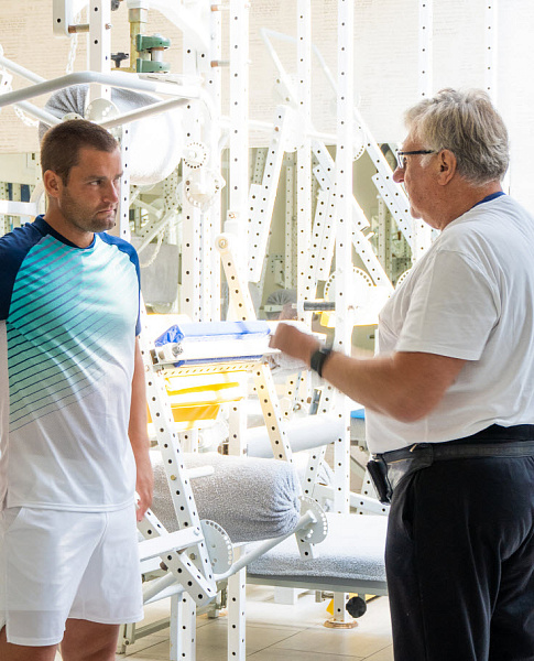
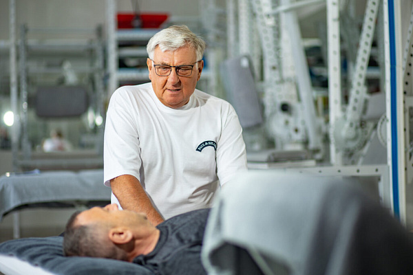
Professor Blum’s approach is distinguished by its detailed analysis of not only the hip joint itself but also the surrounding organs and tissues, their relationships, interdependencies, and interactions. Each segment's role in the development of the disease is assessed. Biomechanical testing is performed using a patented diagnostic system consisting of six macro-blocks, which helps identify the true root cause of the condition. Based on the data obtained, an individualised recovery program is developed with a prognosis of results within specific time frames.
A clear strategy of biomechanical step-by-step progression ensures a stable outcome, even in severe cases of coxarthrosis.
In what types of coxarthrosis is treatment possible?
We accept:
- Patients with grade 1, 2, and 3 coxarthrosis.
- Patients with both primary and secondary hip joint damage.
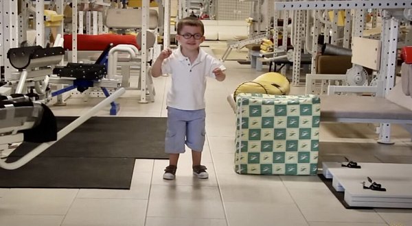
- Infants with congenital hip dysplasia and birth-related hip injuries.
- High-level professional athletes with post-traumatic coxarthrosis.
- Individuals leading a sedentary lifestyle, which can lead to hip joint destruction.
- Women after pregnancy, when increased joint stress has caused disruptions.
- Middle-aged individuals who lead an active lifestyle and seek to prevent coxarthrosis.
We often see patients who have undergone extensive treatment but are unsatisfied with the results. Even in severe cases of coxarthrosis, wh ere hip replacement surgery is recommended, our method can often restore the joint and avoid complex surgery. Our comprehensive, individualised rehabilitation program is developed based on the patient's history, age, disease severity, secondary compensations, and complications.
General description of the problem
Coxarthrosis, or hip joint osteoarthritis, is a progressive disease characterised by the gradual thinning of hyaline cartilage and the growth of bone tissue, leading to joint destruction and loss of mobility. Coxarthrosis is most often the cause of hip pain, with 70% of adults suffering fr om osteoarthritis of the hip joints. Every year, 1.5 million people worldwide undergo hip replacements due to coxarthrosis.
The hip joint is the largest joint in the body, connecting the femur to the pelvis and bearing significant weight. The joint functions like a hinge, with the round head of the femur fitting into the hemispherical acetabulum of the pelvic bone. The surfaces of the bones are covered by strong yet flexible cartilage, which serves as a shock absorber, ensuring smooth movement and protecting the bones from damage.
The blood vessels that nourish the hip joint are closely connected to the gluteal region, the femoral neck, and the round ligament of the femoral head. Therefore, even minor displacements and deformations can disrupt blood flow and impair the joint's tissue nutrition. In most cases, this a biomechanical problem.
As a result, a process of pathological changes begins:
- Disruption of blood circulation in the joint, deterioration of hyaline cartilage nutrition, and reduction of synovial fluid in the joint cavity.
- Impaired circulation of synovial fluid in the joint cavity—cartilage tissue dries out, microcracks appear, and its elasticity and resilience decrease.
- The reduced elasticity leads to a decline in the cartilage’s shock-absorbing function, causing increased friction between the bones. This results in damage to the cartilage, the femoral head, and the surface of the acetabulum of the pelvic bone. The joint becomes damaged, pain occurs, walking becomes difficult, the load is distributed unevenly, and pelvic misalignment worsens.
- Under uneven pressure, the hyaline cartilage thins further, bone tissue proliferates, the joint space narrows, pain intensifies, joint mobility is impaired, and the bones shift. Over time, this leads to an inability to move independently and the necessity of replacing the joint with a prosthesis.
-
Restore the correct biomechanics of the hip joint
-
Eliminate displacements and deformations in the hip joint
-
Correct postural disorders such as pelvic tilt and anterior pelvic tilt
-
Strengthen the ligaments and muscles that stabilize the hip joint
-
Restore the normal distribution of load on the hip joint
-
Improve the mobility and functionality of the joint
-
Prevent further deformations and deterioration of the joint
-
Restore the proper functioning of adjacent organs and tissues affecting the joint's condition
-
Ensure long-term sustainable results and prevent disease recurrence
Objectives of the program
-
Restoring the correct anatomical position of the pelvisRestoring the correct anatomical position of the pelvis, eliminating obliquities, deformations, and displacements of the pelvic and femoral bones, ensuring the proper alignment of both hip joints, and strengthening the muscular-ligamentous structure that stabilises the hip joints.
-
Improving blood circulation and microcirculationImproving blood circulation and microcirculation, creating optimal conditions for the regeneration of the synovial cartilage in the hip joint, and promoting the natural production and circulation of synovial fluid within the joint.
-
Eliminating painEliminating pain, restoring full-range movement in the joints, strengthening and stabilising both hip joints, and returning the patient to an active lifestyle.
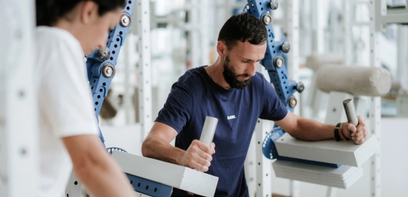
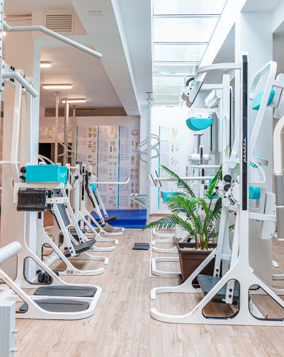
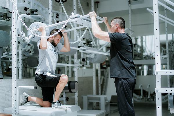
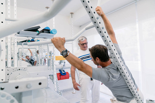
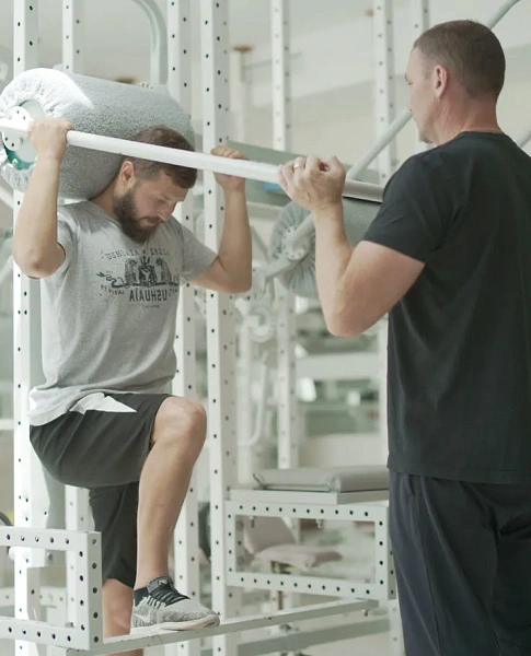
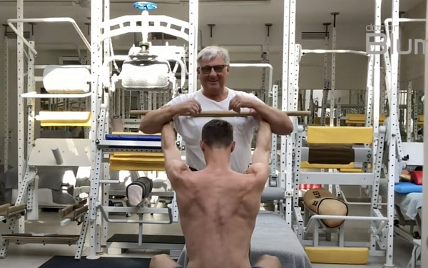
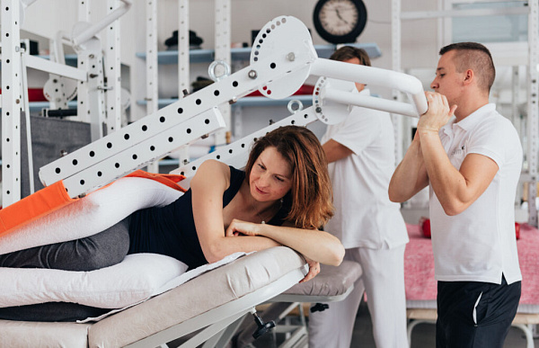
Proprietary methodology
Professor Blum's Centre has been specialising in conservative treatment of coxarthrosis in patients of all ages for many years. The patented system of methods, techniques, and specialised equipment consistently delivers positive results. It alleviates pain, halts the progressive deterioration and destruction of cartilage, restores hip joint mobility, and helps avoid major hip replacement surgery.
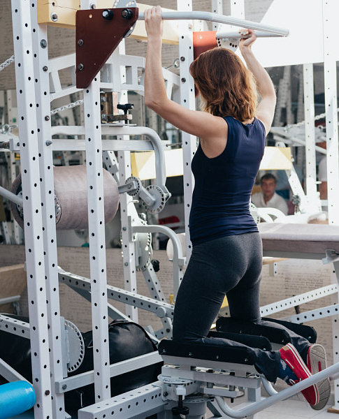
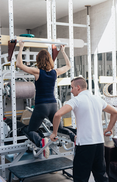
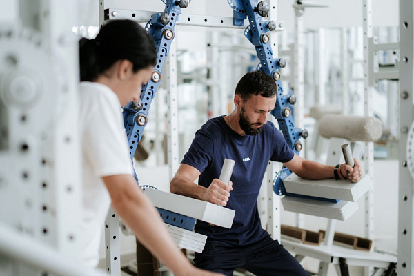
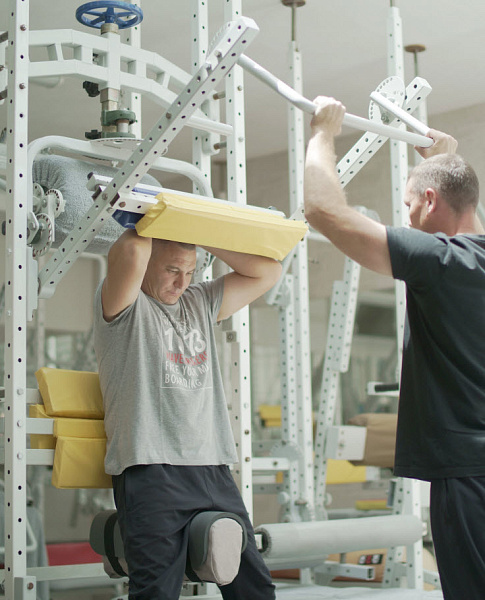
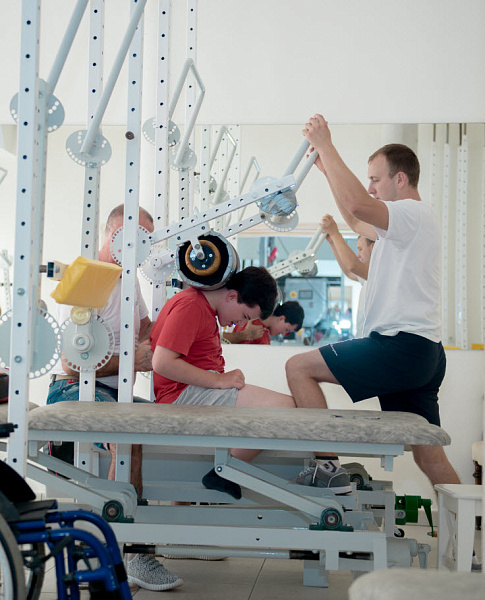
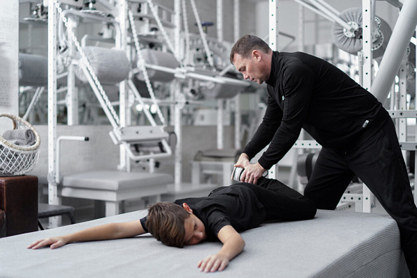
The proprietary rehabilitation equipment offers unparalleled adaptability and precision, with a set range of intensity, specific angles, accelerations, and dozens of other controlled parameters. This tailored approach elicits a targeted response even from the weakest muscular and fascial structures, ensuring the possibility of restoring the hip joint configuration in complex cases.
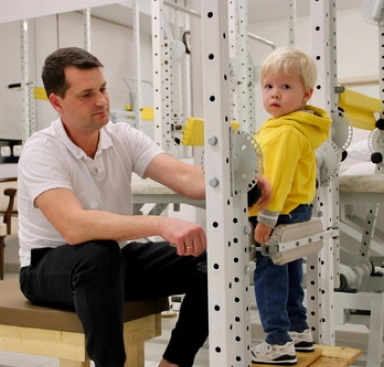
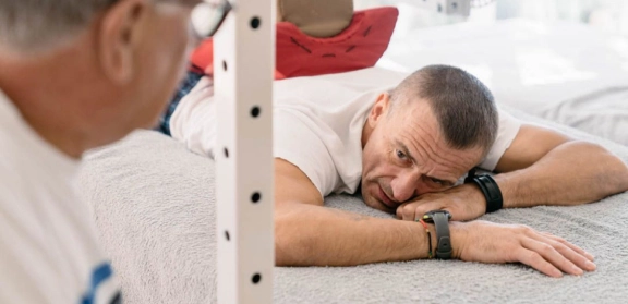
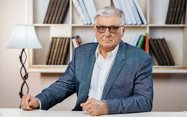
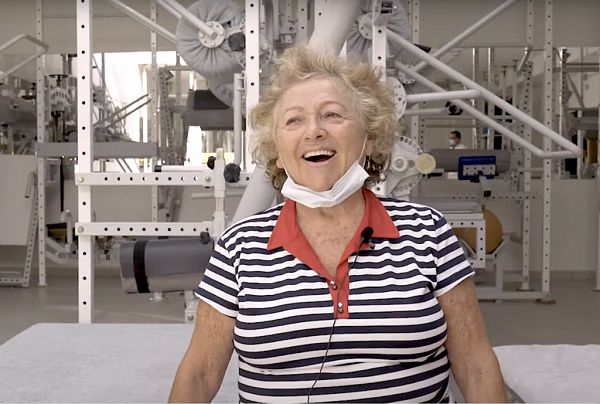
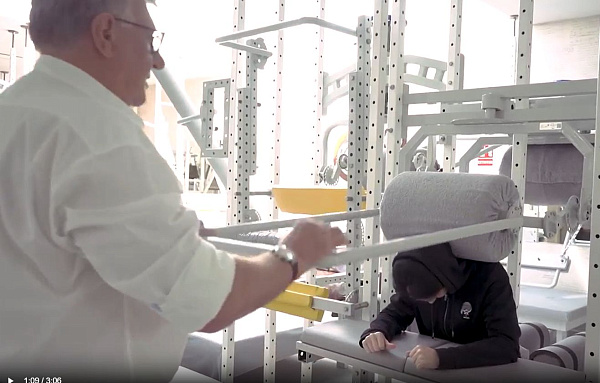
Patient stories

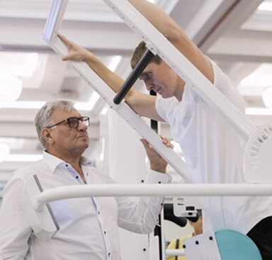
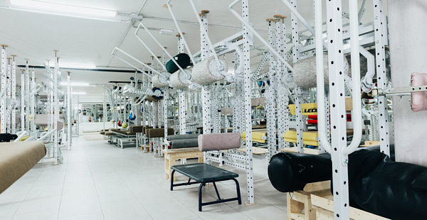
Professor Blum’s Exclusive Rehabilitation System

The center is located in a picturesque corner of the renowned resort town of Marbella, surrounded by cedar trees at the foot of La Concha mountain. Here, science and technology blend with nature, creating a space where the body returns to balance and harmony.
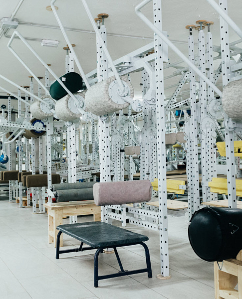
Technologies that deliver results
Select a program
- Personalized Health Recovery Programs
- Disease Prevention
- Customized Check-Up


Q&A
Yes, our Centre has developed specialised rehabilitation programmes for patients after joint replacement surgery. Our goal is to restore maximum range of motion in the joint, balance, and security in walking. However, the most important aspect of rehabilitation after joint replacement, and our unique advantage, is our diagnostic approach, which includes six macro-blocks to identify the underlying cause of joint damage and to develop a plan to address it.
If you experience pain in the hip joints, it is essential to undergo an examination before starting any brisk walking or running. This is to rule out conditions that could worsen with physical activity. It is necessary to take an X-ray of the hip joints, pelvis, and all sections of the spine while standing to get an accurate picture of the joints' condition under load.
The duration of the rehabilitation course depends on the grade of coxarthrosis, the patient's age, and the presence of complications. To determine the exact duration and cost of the course, sign up for a free online consultation with a doctor from the Centre.
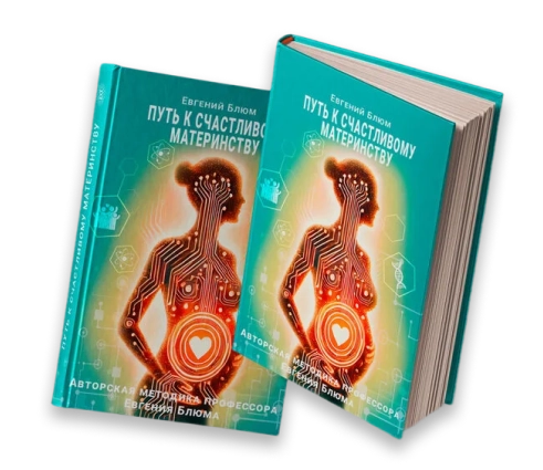
It is necessary to restore the hip joint before pregnancy so that the foetus can grow and develop in a symmetrical body, avoiding the risk of copying deformities from the mother. In addition, an asymmetrical pelvis can affect the organs located in it, potentially causing the uterus to have uneven tone, which is a risk factor for premature labour and caesarean section. Our Centre offers pregnancy preparation programs tailored to identified issues.
Often, symptoms may seem minor, and you can continue living an active life, going to work, while the joint is slowly deteriorating. The symptoms of coxarthrosis often lag behind the actual joint damage. Even with slight clicking, discomfort in the joint, or occasional sharp pain in the hip, an X-ray might already show signs of grade 2 coxarthrosis. Therefore, even with mild symptoms, it is crucial to undergo a full examination to avoid missing the opportunity to restore your joint at an early stage. In your hands you have the opportunity to prevent the need for the surgery. Consult one of our Centre’s specialists for advice.
Coxarthrosis is a progressively worsening condition. Over time, leg pain becomes constant and can even disturb sleep at night. Due to impaired joint stability, the affected leg may shorten, leading to a limp. In severe cases, the person may lose the ability to walk, and it can become painful to sit or even lie down. Hip joint osteoarthritis can eventually lead to ankylosis—complete fusion of the femur with the pelvis—making the leg entirely immobile. This is why it’s essential to begin treatment at the earliest signs of the disease.
At the end of the rehabilitation course, we always provide "homework" — these are constructive recommendations and exercises that will help maintain the achieved results at home.

The cost of a course of treatment with a stay in a hotel
- Appointments and consultations
- Creating an individual program
- Conducting personal sessions
- Appointments and consultations
- Creating an individual program
- Conducting personal sessions
Other areas of work of our Center
.webp)